31 March 2024: Review Articles
Differentiation of Native Vertebral Osteomyelitis: A Comprehensive Review of Imaging Techniques and Future Applications
Weijian Zhu 12BCEF , Sirui Zhou 3D , Jinming Zhang 1D , Li Li 4B , Pin Liu 2A , Wei Xiong 1A*DOI: 10.12659/MSM.943168
Med Sci Monit 2024; 30:e943168
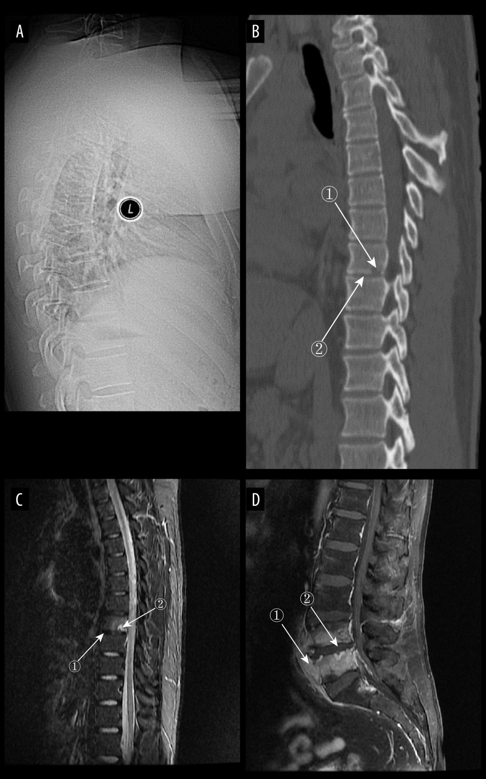
Figure 5 (A) Plain radiograph of early tuberculous spondylitis (TS). No abnormal vertebral changes. (A–C) Images of the same patient, with an interval of 120 days between onset of symptoms and the first radiograph. (B) Plain computed tomography in early TS. There is a less dense lesion with poorly defined borders and no obvious sclerotic bands at marker 1, and an uneroded disc is visible at marker 2. (C) Magnetic resonance imaging (MRI) in early stages of TS. Marker 1 shows that the disc has not received erosion and marker 2 shows an abnormal high signal lesion in the T8 vertebral body. (D) MRI in early stages of TS. Marker 1 shows an abnormally reinforcing shadow in the paravertebral soft tissue, and marker 2 shows relative preservation of the disc, at 120 days between symptom onset and radiograph in this patient (Adobe Illustrator 2022. 26.5. Adobe Inc.).